Sea Stars (Starfish): Anatomically Speaking
Scientists changed the name of the starfish to sea star many years ago. Sea stars don’t look like fish, or swim like fish, and really aren’t fish at all. They belong to the Echinoderm Phylum. Scientists chose the new name, sea star, because sea stars look like a star and live in the sea. But starfish remains a popular name for the sea star to this day.
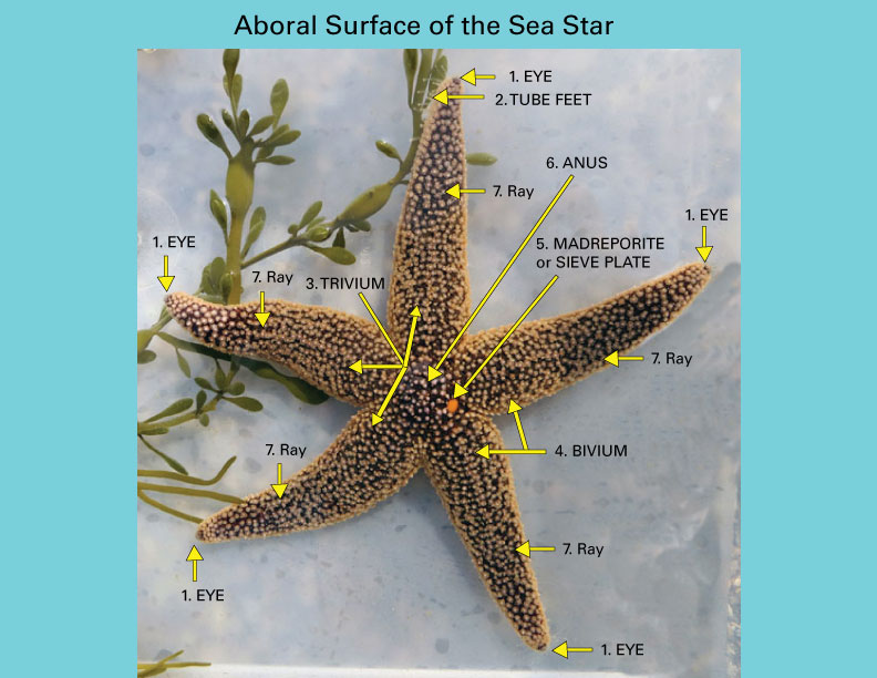
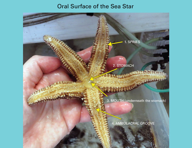
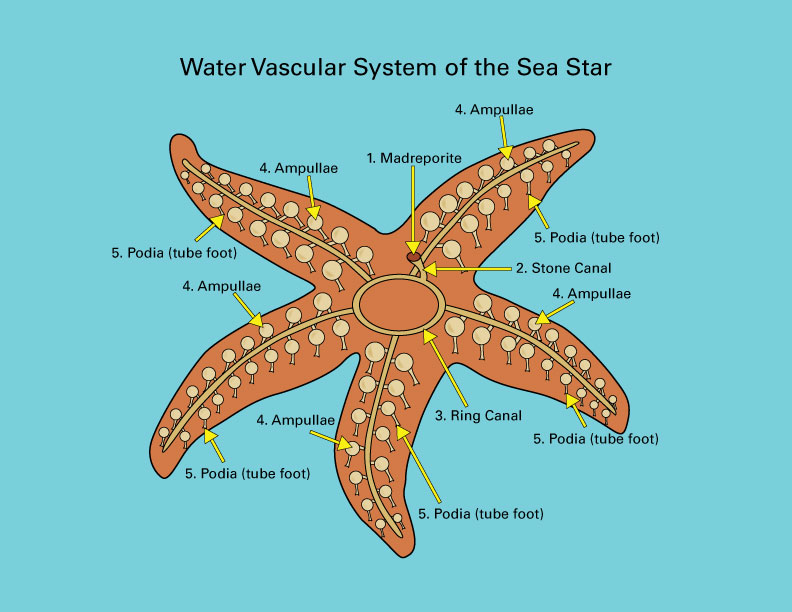
The aboral surface of the sea star, which is the side farthest from the sea stars mouth, is the first image below. The oral surface of the sea star is next, which is the sea star’s underside that’s closest to its mouth. The anatomy visible from both of these surfaces has been identified and defined. The third and final diagram is of the sea star’s water vascular system, that’s extremely important because the sea star uses it to move, eat, breath, and cling to things.

1. Eye: The common sea star has five eye spots on the tip of each of its five rays. These eye spots can see shadows and light.
2. Tube feet: Sometimes called podia, the sea star’s tube feet extend from the underside of each of the sea star’s rays. Tube feet can be visible from the aboral surface as they stretch out to move the sea star from one location to another. Tube feet aren’t only used for locomotion, they’re also used to hold onto food and pry clam shells and other mollusks open. The surface of the tube feet can exchange gases and nitrogen waste.
3. Trivium: The three rays that are farthest from the sea star’s madreporite.
4. Bivium: The two rays closest to the sea star’s madreporite.
5. Madreporite or sieve plate: This is the reddish-orange, or sometimes white spot towards the center, top of the sea star’s body that lets water into it’s water vascular system.
6. Anus: The end of the digestive tract where waste is ejected, although most undigested food is regurgitated rather than released through the sea star’s anus.
7. Rays: Common sea stars have five rays, unless they lose one or grow an extra. Most sea stars have 5-14 rays, but sunflower sea stars can have up to 15-24 rays. If a sea star loses a ray to a predator or an accident, it can grow a new one back again. The process of growing a new ray is called regeneration.

1. Spines: The sea star’s surface has many white spines that give the sea star a rough feel, and are used for protection.
2. Stomach: A sea star’s able to eat its prey outside its body by dropping its cardiac stomach, which looks and feels like an egg white, out of its mouth and into its prey’s shells. The stomach wraps around its prey and digests it outside of its body. Then the sea star pulls its stomach back into its body through its mouth. A sea star’s favorite food is shellfish, like mussels, clams, and barnacles.
3. Mouth: The sea star’s mouth is located in the center of its body, underneath. Part of the sea star’s stomach connects to its mouth, and when there’s food available, the sea star’s stomach emerges from its mouth to eat.
4. Ambulacral Groove: This is the area that contains the sea star’s tube feet. It’s located underneath each ray.

1. Madreporite or sieve plate: a small, smooth plate, at the entrance of the sea star’s water vascular system, through which the sea star takes in sea water. It’s located on the aboral side of the sea star, slightly off the center.
2. Stone Canal: a tube connecting the sea star’s madreporite to its ring canal that’s the second part of the sea star’s water vascular system.
3. Ring Canal: the circular tube of the sea star’s water vascular system that connects the stone canal to the ampullae in its rays.
4. Ampullae: A pouch or sack-like part of the sea star’s water vascular system that expands and contracts to move water up and down each tube foot. When the sea star wants to create a suction at the end of its tube foot, its ampullae pulls water out of the podia. When releasing the suction, the ampullae pushes water into the end of each tube foot.
5. Podia (tube foot): A podia or tube foot is one of the small, flexible extensions of the sea star’s water vascular system that has a suction cup at the end. This suction cup allows the sea star to hold tightly to rocks and shells of its prey. Tube feet also help the sea star to move, and the podia’s surface can exchange gases and nitrogen waste. Tube feet are located on the underside of the sea star’s ray, in the ambulacral groove.
